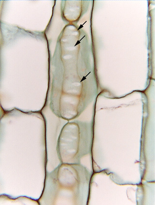
 Fig. 3.1-3.
Longitudinal section of stem of milkweed (Asclepias). This micrograph
shows three columns of parenchyma cells; those on the left and right have been
cut down the center, so their front and back walls are missing, just as in Fig.
3.1-1. But the middle row of cells has been cut just where those cells are
pressed up against and contact another row of cells close to us. The vertically
elongate ovals, outlined in material stained brown, are the contact
faces: the
point where two cells press against each other is the contact face. If both
cells are spheres, the contact face is round, but if the two cells are columnar,
the contact face is oval, as are these in the micrograph. If the cells barely
touch each other, the contact faces are small, but if the two cells are forced
firmly against each other, the contact face is larger. Within each contact face
are numerous smaller oval areas (arrows), so pale they appear white. They look like holes
in the wall but are actually primary pit fields -- areas where the two primary
walls of the contact face are unusually thin and plasmodesmata occur in high
density (the plasmodesmata cannot be seen by ordinary light microscopy). Examine
Fig. 3.1-1: those cells fit together so tightly that all
the walls are contact faces except the small regions that face the intercellular
spaces. There are almost certainly many primary pit fields present in the walls
of Fig. 3.1-1, but they cannot be seen in the micrograph. Fig.
3.1-2 is somewhat surprising in having such a large expanse of primary wall
without having any primary pit fields, but it might be that all of that wall
faces an intercellular space -- such wall do not have primary pit fields.
Fig. 3.1-3.
Longitudinal section of stem of milkweed (Asclepias). This micrograph
shows three columns of parenchyma cells; those on the left and right have been
cut down the center, so their front and back walls are missing, just as in Fig.
3.1-1. But the middle row of cells has been cut just where those cells are
pressed up against and contact another row of cells close to us. The vertically
elongate ovals, outlined in material stained brown, are the contact
faces: the
point where two cells press against each other is the contact face. If both
cells are spheres, the contact face is round, but if the two cells are columnar,
the contact face is oval, as are these in the micrograph. If the cells barely
touch each other, the contact faces are small, but if the two cells are forced
firmly against each other, the contact face is larger. Within each contact face
are numerous smaller oval areas (arrows), so pale they appear white. They look like holes
in the wall but are actually primary pit fields -- areas where the two primary
walls of the contact face are unusually thin and plasmodesmata occur in high
density (the plasmodesmata cannot be seen by ordinary light microscopy). Examine
Fig. 3.1-1: those cells fit together so tightly that all
the walls are contact faces except the small regions that face the intercellular
spaces. There are almost certainly many primary pit fields present in the walls
of Fig. 3.1-1, but they cannot be seen in the micrograph. Fig.
3.1-2 is somewhat surprising in having such a large expanse of primary wall
without having any primary pit fields, but it might be that all of that wall
faces an intercellular space -- such wall do not have primary pit fields.