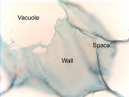
 Fig. 3.1-2. Transverse section of stem pith in beet (Beta
vulgaris). This is a high magnification view in which part of the front
wall of a cell is present, it is the sheet of bluish material. On the
upper left, the microtome knife passed through the wall and cut part of it away,
leaving just a view of the vacuole (white area). The remaining sheet of front
wall is so delicate that it has folded and wrinkled slightly, probably during
the agitation that occurs as the slide was being stained. This wall is
remarkably smooth and uniform; it has no primary pit fields and although there
must be cytoplasm lying against this wall, pink-stained plastids are visible.
There is a triangular intercellular space surrounded by bluish material which is
the walls of the three cells that meet at the space. In Fig.
3.1-1, the cell walls come straight upward at us so the knife cut them
cleanly and the walls look thin, but here each wall curves strongly so the knife
has cut each at least partially in face view.
Fig. 3.1-2. Transverse section of stem pith in beet (Beta
vulgaris). This is a high magnification view in which part of the front
wall of a cell is present, it is the sheet of bluish material. On the
upper left, the microtome knife passed through the wall and cut part of it away,
leaving just a view of the vacuole (white area). The remaining sheet of front
wall is so delicate that it has folded and wrinkled slightly, probably during
the agitation that occurs as the slide was being stained. This wall is
remarkably smooth and uniform; it has no primary pit fields and although there
must be cytoplasm lying against this wall, pink-stained plastids are visible.
There is a triangular intercellular space surrounded by bluish material which is
the walls of the three cells that meet at the space. In Fig.
3.1-1, the cell walls come straight upward at us so the knife cut them
cleanly and the walls look thin, but here each wall curves strongly so the knife
has cut each at least partially in face view.