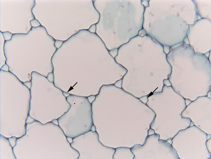
 Fig.
3.1-1. Transverse section of pith in ragweed stem (Ambrosia).
The large irregular objects that appear empty are parenchyma
cells, the small areas are intercellular spaces (arrows), and the dark lines
that make up the image are primary cell walls. Parenchyma cells in pith are
often some of the largest cells in any plant, being so large that during the
cutting of the section, most of every cell is cut away: These walls appear to be
clean and distinct because they come straight up at us -- both the back and
front walls of each cell have been cut off. These cells appear empty because
each consists almost entirely of central vacuole, and during preparation for
microscopy, the vacuole contents leaked out and were washed away -- at this
point, the cells really are virtually empty. Because each central vacuole is so
large, its vacuole membrane is pushed close to the cell's plasma membrane, which
itself is pressed against the wall. There is only a very small amount of
cytoplasm, squeezed into such a thin, flat layer near the wall that it is almost
impossible to see, even at high power. Several cells here have a faint bluish
material in them, that is the protoplasm in face view: the section has just
grazed either the front or back wall and the thin layer of cytoplasm. Being so
thin, it has absorbed only a little stain, so it appears faint, with a few pink
dots that are probably plastids. Nuclei would be large enough to be visible at
this magnification, but none is present just due to luck -- in each of the cells
here, the nucleus must have been either in front of or behind the cuts that made
this section, so the nuclei were cut away.
Fig.
3.1-1. Transverse section of pith in ragweed stem (Ambrosia).
The large irregular objects that appear empty are parenchyma
cells, the small areas are intercellular spaces (arrows), and the dark lines
that make up the image are primary cell walls. Parenchyma cells in pith are
often some of the largest cells in any plant, being so large that during the
cutting of the section, most of every cell is cut away: These walls appear to be
clean and distinct because they come straight up at us -- both the back and
front walls of each cell have been cut off. These cells appear empty because
each consists almost entirely of central vacuole, and during preparation for
microscopy, the vacuole contents leaked out and were washed away -- at this
point, the cells really are virtually empty. Because each central vacuole is so
large, its vacuole membrane is pushed close to the cell's plasma membrane, which
itself is pressed against the wall. There is only a very small amount of
cytoplasm, squeezed into such a thin, flat layer near the wall that it is almost
impossible to see, even at high power. Several cells here have a faint bluish
material in them, that is the protoplasm in face view: the section has just
grazed either the front or back wall and the thin layer of cytoplasm. Being so
thin, it has absorbed only a little stain, so it appears faint, with a few pink
dots that are probably plastids. Nuclei would be large enough to be visible at
this magnification, but none is present just due to luck -- in each of the cells
here, the nucleus must have been either in front of or behind the cuts that made
this section, so the nuclei were cut away.
Notice that the intercellular spaces are all triangular, indicating that each is formed where three cells meet; it is extremely rare for four cells to meet at one point.
Related pages in Plant Anatomy (Mauseth): 43-44.