|
[Home]
[Up]
[Ordinary epidermis]
[Guard cells]
[Thick cuticle]
[Thicker cuticle]
[Thin cuticle]
[Parasitic plant]
[Petal epidermis]
[Sclerified epidermis]
[Papillose epidermis]
[Sculptured cuticle]
[Elaborate cuticle]
[Cuticular horns]
[Radial walls]
[Cuticle proper]
[No epidermis]
[Epidermal peels]
[Cycad peel]
[Paradermal]
[Typical stoma]
[Sunken stoma]
[Stomatal orientation 1]
[Unusual orientation 2]
[Artifact]
[Stomata and fibers]
[Stomatal crypts]
[Crypts, mag.]
[Crypt margin]
[Non-crypt]
[Water lily]
[Stomatal channels]
[Groove, hi mag]
[Subsidiary cells]
[Ledges]
[Papillae]
[Trichome]
[Uniseriate hair]
[Peltate hair, mag]
[Peltate, lo mag]
[Branched hairs]
[Trichome base]
[Lithocyst, Ficus]
[Lithocysts, hemp]
[Bulliform cells]
[Grass epidermis]
[Multiple epi]
[Uniseriate?]
[Peperomia]
| |
 Chapter
10. Epidermis. Chapter
10. Epidermis.
The figures presented here were selected to illustrate aspects of the
epidermis and the many types of cells that occur as part of the epidermis. More
than many other tissues, the epidermis may be extremely complex in some plants;
the epidermis of stems and leaves may differ from that of the roots, of course,
but also may differ from the epidermis of sepals, petals, stamens, and carpels.
Even the two sides of a single leaf typically differ significantly.
Consequently, this chapter is longer than many of the others, with micrographs
illustrating a wider diversity of cell types and arrangements.
As
in the other chapters, these legends have not been written to be a complete
discussion of all aspects of the epidermis, but rather to complement the
more complete discussion of the epidermis presented in Chapter104 (pages 167 to
198)
in the textbook Plant Anatomy by J. D. Mauseth, published by Cummings
& Hathaway.
Click here for a set of links to the micrographs of this
chapter.
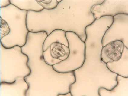
Epidermal peel, showing
ordinary epidermal cells and a stomatal complex.
|