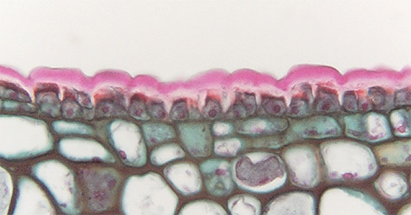
 Fig.
10.2-14. Transverse section of Ficus leaf (related to fig).
This is an
extremely thick cuticle proper (the pink layer) and cutinized wall (the white
layer). The shape of the protoplasts is not the shape of the cell:
the whitish areas are cell walls with cutin deposited in them, and their shape
indicates that the outer parts of the radial walls are thickened, as is all of
the outer wall. After secreting enough cutin to encrust all this wall, the cells
continued to produce enough cutin to build a thick layer above the cells.
Fig.
10.2-14. Transverse section of Ficus leaf (related to fig).
This is an
extremely thick cuticle proper (the pink layer) and cutinized wall (the white
layer). The shape of the protoplasts is not the shape of the cell:
the whitish areas are cell walls with cutin deposited in them, and their shape
indicates that the outer parts of the radial walls are thickened, as is all of
the outer wall. After secreting enough cutin to encrust all this wall, the cells
continued to produce enough cutin to build a thick layer above the cells.