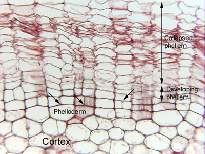
 Fig.
17.1-1. Transverse section of geranium
stem (Pelargonium). When a geranium stem develops a tan color instead of
green, it has begun producing cork. This micrograph shows the outer parts of the
cortex, covered in a bark that consists of many layers of phellem (cork cells).
Cork cells are often empty and dead at maturity and they often collapse, like
these. Notice that the two arrows on the left point to sites where two rows of
phellem cells meet a single large cell: the single large cell cannot be
producing two rows, so it is not the phellogen (the cork cambium). The pairs of small cells at the arrow tips must be
phellogen cells. Other cells at the same level are also cork cambium
cells, but it is only where two rows meet one cell that cork cambium cells are
easy to identify. The right arrow points to a pair of cells that underlies just
a single row of cork cells: a cork cambium cell has just undergone a division
into two cambium cells, and if the sample had not been dissected for
examination, the pair would have begun producing two rows of cork cells rather
than just the one row.
Fig.
17.1-1. Transverse section of geranium
stem (Pelargonium). When a geranium stem develops a tan color instead of
green, it has begun producing cork. This micrograph shows the outer parts of the
cortex, covered in a bark that consists of many layers of phellem (cork cells).
Cork cells are often empty and dead at maturity and they often collapse, like
these. Notice that the two arrows on the left point to sites where two rows of
phellem cells meet a single large cell: the single large cell cannot be
producing two rows, so it is not the phellogen (the cork cambium). The pairs of small cells at the arrow tips must be
phellogen cells. Other cells at the same level are also cork cambium
cells, but it is only where two rows meet one cell that cork cambium cells are
easy to identify. The right arrow points to a pair of cells that underlies just
a single row of cork cells: a cork cambium cell has just undergone a division
into two cambium cells, and if the sample had not been dissected for
examination, the pair would have begun producing two rows of cork cells rather
than just the one row.
The larger cells just below the cork cambium resemble cortex cells, but they have flattened walls where they are in contact with the cork cambium cells. These are phelloderm cells.