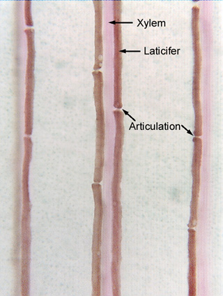
 Fig.
9.2-4. Clearing of banana leaf (Musa). The
three vertical strips of tissue are leaf vascular bundles, with the pink color
being xylem and the brown being latex. The leaf was cleared by placing large
pieces of it in warm potassium hydroxide and a stain solution. Latex tubes
typically appear empty in histological slides because the latex drains out of
the cut ends of the long tube-like cells. But these are articulated
laticifers – a structure made up of many cells. The articulations
are visible between the individual cells because the brown color is due to
stained latex, and the latex is excluded from the area between cells by the
presence of the cells’ end walls (which did not stain). There is a narrow bit
of brown color between some cells – apparently the end walls have a small hole
or two that lets latex move from cell to cell.
Fig.
9.2-4. Clearing of banana leaf (Musa). The
three vertical strips of tissue are leaf vascular bundles, with the pink color
being xylem and the brown being latex. The leaf was cleared by placing large
pieces of it in warm potassium hydroxide and a stain solution. Latex tubes
typically appear empty in histological slides because the latex drains out of
the cut ends of the long tube-like cells. But these are articulated
laticifers – a structure made up of many cells. The articulations
are visible between the individual cells because the brown color is due to
stained latex, and the latex is excluded from the area between cells by the
presence of the cells’ end walls (which did not stain). There is a narrow bit
of brown color between some cells – apparently the end walls have a small hole
or two that lets latex move from cell to cell.
The laticifers run parallel to the vascular bundles, but just below them; when the microscope is focused on the laticifers, the xylem is so far above the plane of focus that its red-stained helical secondary walls merely look like the pink strip we see here. If we focused up to see the xylem, the laticifers would be indistinct.