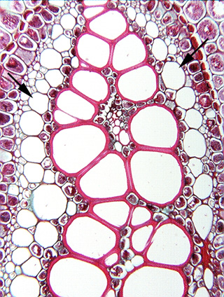
 Fig.
8.2-9. Transverse section of a fern rhizome (Pteridium
aquilinum). The center of this micrograph is occupied by xylem (the large
red-stained cells), and surrounding that is a band of large, empty-looking cells
with thin walls (arrows); those are the sieve cells
of this fern phloem.
Fig.
8.2-9. Transverse section of a fern rhizome (Pteridium
aquilinum). The center of this micrograph is occupied by xylem (the large
red-stained cells), and surrounding that is a band of large, empty-looking cells
with thin walls (arrows); those are the sieve cells
of this fern phloem.