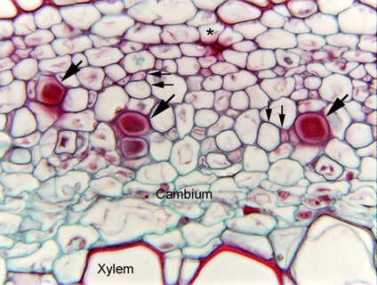
 Fig.
8.1-3. Transverse section of ragweed stem (Ambrosia).
Ragweed is a dicot with phloem that is relatively easy to study. The two pairs
of small arrows indicate pairs of sieve tube members and companion cells. The
three large arrows point to dark red-stained areas – those are P-protein
plugs, the masses of protein that become trapped at the sieve plate
of damaged sieve tube members, sealing them and preventing the phloem sap from
leaking out. Here, these are artifacts
caused when the phloem was damaged during dissection. If the tissue had been
fixed very gently, the plugs would not have formed; however, it is actually
quite handy to have them for studying phloem – they indicate where actively
conducting phloem is located, and since they form at the sieve plate, we know
that four sieve plates must be located at about the same level here. The other
sieve tube members in this sample probably also have P-protein plugs but they
look empty because the sieve plate and the plug are located so much higher or
lower that this section missed them.
Fig.
8.1-3. Transverse section of ragweed stem (Ambrosia).
Ragweed is a dicot with phloem that is relatively easy to study. The two pairs
of small arrows indicate pairs of sieve tube members and companion cells. The
three large arrows point to dark red-stained areas – those are P-protein
plugs, the masses of protein that become trapped at the sieve plate
of damaged sieve tube members, sealing them and preventing the phloem sap from
leaking out. Here, these are artifacts
caused when the phloem was damaged during dissection. If the tissue had been
fixed very gently, the plugs would not have formed; however, it is actually
quite handy to have them for studying phloem – they indicate where actively
conducting phloem is located, and since they form at the sieve plate, we know
that four sieve plates must be located at about the same level here. The other
sieve tube members in this sample probably also have P-protein plugs but they
look empty because the sieve plate and the plug are located so much higher or
lower that this section missed them.
Notice the dark, irregular mass just below the asterisk near the top of the micrograph: that is probably a collapsed sieve tube member of the protophloem.