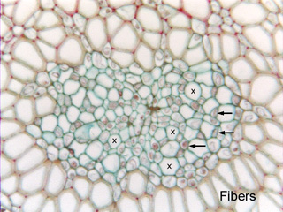
 Fig.
8.1-1. Transverse section of stem of Cordyline
(a monocot related to agaves). Monocots are favorite plants for introducing
students to phloem because monocot
phloem has a regular pattern of large, empty-looking sieve tube members and
small, cytoplasmic companion cells. Because it is rare to ever
encounter any other cell type in monocot phloem, it is relatively easy to see
these two important cell types. In contrast, the phloem of dicots can be a
mixture of many things, and dicot sieve tube members are often narrow;
consequently, most species of dicots are poor teaching material as far a phloem
is concerned.
Fig.
8.1-1. Transverse section of stem of Cordyline
(a monocot related to agaves). Monocots are favorite plants for introducing
students to phloem because monocot
phloem has a regular pattern of large, empty-looking sieve tube members and
small, cytoplasmic companion cells. Because it is rare to ever
encounter any other cell type in monocot phloem, it is relatively easy to see
these two important cell types. In contrast, the phloem of dicots can be a
mixture of many things, and dicot sieve tube members are often narrow;
consequently, most species of dicots are poor teaching material as far a phloem
is concerned.
This vascular bundle has a typical monocot arrangement of sieve tube members (several are marked by X) and companion cells (some marked by arrows). The dark material in the companion cells is nucleus and cytoplasm – remember the sieve tube members lose their nuclei during maturation, and companion cell nuclei must exert control over both the companion cell and the associated sieve tube member. Sieve tube members typically appear clean and white, with no contents, no plasmolysis. In life – before they were dissected and fixed – they have only a thin film of cytoplasm bound firmly to the wall. It usually stays in place during fixation, giving the walls a slightly thickened appearance (the cytoplasm often stains the same color as the wall).
This phloem is surrounded by a thick sheath of fibers.