 Fig.
7.3-4. Transverse section of vascular bundle in parsnip (Pastinaca).
This micrograph shows a simple perforation plate lying parallel to the plane of
sectioning, but it is in a vessel element with a helical secondary wall rather
than a pitted one as in Fig. 7.3-3. The
arrow marks the portion of wall that is the perforation plate, the other red
bands are helices of secondary wall.
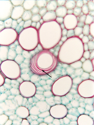
Fig.
7.3-4. Transverse section of vascular bundle in parsnip (Pastinaca).
This micrograph shows a simple perforation plate lying parallel to the plane of
sectioning, but it is in a vessel element with a helical secondary wall rather
than a pitted one as in Fig. 7.3-3. The
arrow marks the portion of wall that is the perforation plate, the other red
bands are helices of secondary wall.