 Fig.
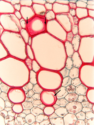
7.3-1. Transverse section of stem of sweet potato
(Ipomoea batatas). In the upper part of the micrograph, a perforation
plate was lying parallel to the plane of the section, so we see it in face view.
The entire red area from arrow to arrow is the perforation plate, the hole in
the center is the perforation itself. Because there is just the one hole in the
plate, it is a simple
perforation plate rather than a compound perforation plate. When the
cell was still developing, this end wall was complete and then would have been
called a perforation partition. In the last stages of differentiation, the
center of the perforation partition was digested away, creating the perforation.
The remaining wall is the rim; this rim is unusually wide -- typically they are
much less than half this wide and the perforation occupies more of the
perforation plate. Very often, the perforation plate is tilted, so in a section
like this, part of it is cut away and what is left appears as just an arc of
wall material.
Fig.
7.3-1. Transverse section of stem of sweet potato
(Ipomoea batatas). In the upper part of the micrograph, a perforation
plate was lying parallel to the plane of the section, so we see it in face view.
The entire red area from arrow to arrow is the perforation plate, the hole in
the center is the perforation itself. Because there is just the one hole in the
plate, it is a simple
perforation plate rather than a compound perforation plate. When the
cell was still developing, this end wall was complete and then would have been
called a perforation partition. In the last stages of differentiation, the
center of the perforation partition was digested away, creating the perforation.
The remaining wall is the rim; this rim is unusually wide -- typically they are
much less than half this wide and the perforation occupies more of the
perforation plate. Very often, the perforation plate is tilted, so in a section
like this, part of it is cut away and what is left appears as just an arc of
wall material.
Notice that all the vessels in the upper part of the micrograph have the beaded wall that indicates they have circular bordered pits. In the lower part of the micrograph, the arrow indicates a vessel with a fragment of red wall material in it -- that is a vessel element with helical secondary walls, and during microtoming, the knife broke it loose from the primary wall.