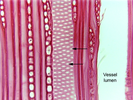
 Fig.
7.2-7. Longitudinal section of wood of American
hornbeam (Carpinus caroliniana), a dicot. The two arrows point out circular
bordered pits in the side wall of a vessel element. There is no torus
or margo; instead, the white dot in the center of each pit is where we are
looking through the pit aperture. The gray region around each aperture is the
border – we are looking through the area where the secondary wall is lifted up
away from the primary wall. It looks gray because of the amount of light coming
through that region – it is actually stained red. The dark red lines that
outline the shape of each circular bordered pit is the region where the
secondary walls are firmly attached to the primary walls. Although these are
properly called “circular bordered pits,” they are actually hexagonal in
shape; however, there is no such term as “hexagonal bordered pits.” For
every pit we see in this vessel element, there is a corresponding pit in the
vessel element behind it. Rather than just pits, we are seeing the front half of
pit-pairs.
Notice just how porous these walls are – there are many, many sites at which water can pass from one tracheary element to another. But keep in mind that these are not complete holes – there is a pit membrane (the two primary walls and middle lamella) separating each pit of the pit pair. That pit membrane reduces the likelihood of any air bubble passing from one cell to another.