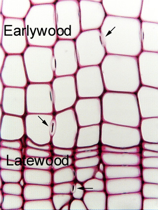
 Fig.
7.1-6. Transverse section of pine wood (Pinus).
All cells in this micrograph are tracheids.
You can not tell that from simply looking at this section, but rather by knowing
that it is a sample of pine, which has lacks both vessels and fibers. The three
arrows indicate circular bordered pits; any
cell with bordered pits will be a tracheary element, not a fiber. If
this were an unknown sample, you could search for perforations -- if these were
vessels, a perforation should be visible somewhere (although in transverse
section, it might be difficult to recognize). An
important clue that these are tracheids and not vessels is that they
are all so similar in diameter -- they are uniformly large in the earlywood and
uniformly smaller in the latewood. Vessels typically are present in several
sizes, and are virtually always associated with other cells that are smaller
than they. The uniformity here, along with the circular bordered pits, indicates
that these are tracheids.
Fig.
7.1-6. Transverse section of pine wood (Pinus).
All cells in this micrograph are tracheids.
You can not tell that from simply looking at this section, but rather by knowing
that it is a sample of pine, which has lacks both vessels and fibers. The three
arrows indicate circular bordered pits; any
cell with bordered pits will be a tracheary element, not a fiber. If
this were an unknown sample, you could search for perforations -- if these were
vessels, a perforation should be visible somewhere (although in transverse
section, it might be difficult to recognize). An
important clue that these are tracheids and not vessels is that they
are all so similar in diameter -- they are uniformly large in the earlywood and
uniformly smaller in the latewood. Vessels typically are present in several
sizes, and are virtually always associated with other cells that are smaller
than they. The uniformity here, along with the circular bordered pits, indicates
that these are tracheids.