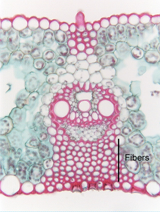
 Fig.
5.1-1. Transverse section of a grass leaf (Poa). This is a
transverse section through a vascular bundle and a mass of sclerenchyma cells
– fibers, in this case – that runs the
length of the leaf. Although the sclerenchyma cells appear spherical here, in
longitudinal section, they would be very long. These cells have both primary and
secondary walls that contain lignin, so the dye Safranin has stained them red.
The cells on the sides of the micrograph are parenchyma cells with unlignified
primary walls, so despite being exposed to Safranin, they have stained green due
to the presence of Fast Green in the Safranin/Fast Green staining procedure. The
epidermis cells too have stained red, almost certainly because they have cutin
in their walls, rather than lignin (Safranin is not a specific stain that reacts
with only one chemical). The large cells in the vascular bundle are vessel
elements, xylem cells that conduct water; they are a type of conducting
sclerenchyma.
Fig.
5.1-1. Transverse section of a grass leaf (Poa). This is a
transverse section through a vascular bundle and a mass of sclerenchyma cells
– fibers, in this case – that runs the
length of the leaf. Although the sclerenchyma cells appear spherical here, in
longitudinal section, they would be very long. These cells have both primary and
secondary walls that contain lignin, so the dye Safranin has stained them red.
The cells on the sides of the micrograph are parenchyma cells with unlignified
primary walls, so despite being exposed to Safranin, they have stained green due
to the presence of Fast Green in the Safranin/Fast Green staining procedure. The
epidermis cells too have stained red, almost certainly because they have cutin
in their walls, rather than lignin (Safranin is not a specific stain that reacts
with only one chemical). The large cells in the vascular bundle are vessel
elements, xylem cells that conduct water; they are a type of conducting
sclerenchyma.