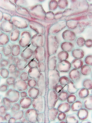
 Fig.
3.5-3. Paradermal section through the leaf of ground ivy (Glecoma,
in the mint family, not a real ivy). The long strands with helical red bands are
xylem cells that conduct water. At this point, focus on the larger, rounded
parenchyma cells that touch the conducting cells (arrows indicate three of
many). The conducting cells are
slightly scalloped because the parenchyma cells pressed so firmly against them
while they were maturing. The dark material in all the parenchyma cells are
chloroplasts packed so closely that it is difficult to tell that they are
individual bean-shaped organelles. In parenchyma cells that contact xylem
conducting cells, chloroplasts are located along the walls away from the
conducting cell. This leaf – like most material used for general studies of
plant anatomy – was prepared by embedding it in wax and then cutting it into
sections about 10 to 12mm
thick. This is too thick to be able to see labyrinthine walls, but almost
certainly, the parenchyma cell walls that touch the xylem conducting cells are transfer
walls with labyrinthine walls. To be certain, it is necessary to use
either scanning or transmission electron microscopy. If these cells do indeed
have labyrinthine walls, then they are transfer cells.
Fig.
3.5-3. Paradermal section through the leaf of ground ivy (Glecoma,
in the mint family, not a real ivy). The long strands with helical red bands are
xylem cells that conduct water. At this point, focus on the larger, rounded
parenchyma cells that touch the conducting cells (arrows indicate three of
many). The conducting cells are
slightly scalloped because the parenchyma cells pressed so firmly against them
while they were maturing. The dark material in all the parenchyma cells are
chloroplasts packed so closely that it is difficult to tell that they are
individual bean-shaped organelles. In parenchyma cells that contact xylem
conducting cells, chloroplasts are located along the walls away from the
conducting cell. This leaf – like most material used for general studies of
plant anatomy – was prepared by embedding it in wax and then cutting it into
sections about 10 to 12mm
thick. This is too thick to be able to see labyrinthine walls, but almost
certainly, the parenchyma cell walls that touch the xylem conducting cells are transfer
walls with labyrinthine walls. To be certain, it is necessary to use
either scanning or transmission electron microscopy. If these cells do indeed
have labyrinthine walls, then they are transfer cells.