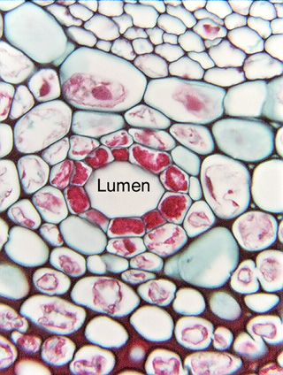
 Fig. 3.2-6.
Transverse section of wormwood (Artemisia). This circle of dark red cells
represents a transverse section of a long, tube-like secretory
duct: the entire tube consists of these densely cytoplasmic cells
that secrete their product into the duct lumen, the white "empty"
space (empty only because the secretory product was washed out as the tissue was
processed). The red material in each cell is all the cell's protoplasm, not just
its nucleus; even though the red material does look like a lumpy nucleus, it is
just too abundant and too lumpy to actually be a nucleus. The red material is
also quite abundant just for protoplasm, but secretory parenchyma cells are
often densely cytoplasmic with little vacuolation. Only a single layer appears
to be secretory, as all surrounding parenchyma cells have protoplasm that has
stained less intensely (they are also extremely plasmolyzed -- the fixative
probably had too much alcohol in it so it absorbed water from the cells). Cells
on the top of the micrograph have thicker walls -- these are collenchyma cells,
discussed in Chapter 4 (pages 53 to 59 in Plant Anatomy (Mauseth)).
Fig. 3.2-6.
Transverse section of wormwood (Artemisia). This circle of dark red cells
represents a transverse section of a long, tube-like secretory
duct: the entire tube consists of these densely cytoplasmic cells
that secrete their product into the duct lumen, the white "empty"
space (empty only because the secretory product was washed out as the tissue was
processed). The red material in each cell is all the cell's protoplasm, not just
its nucleus; even though the red material does look like a lumpy nucleus, it is
just too abundant and too lumpy to actually be a nucleus. The red material is
also quite abundant just for protoplasm, but secretory parenchyma cells are
often densely cytoplasmic with little vacuolation. Only a single layer appears
to be secretory, as all surrounding parenchyma cells have protoplasm that has
stained less intensely (they are also extremely plasmolyzed -- the fixative
probably had too much alcohol in it so it absorbed water from the cells). Cells
on the top of the micrograph have thicker walls -- these are collenchyma cells,
discussed in Chapter 4 (pages 53 to 59 in Plant Anatomy (Mauseth)).