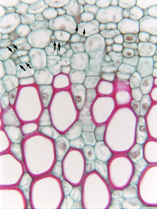
 Fig.
11.5-8. Transverse section of pea stem (Pisum sativum). This
micrograph shows the metaxylem and metaphloem of a vascular bundle. Notice that parenchyma makes up a large fraction
of the xylem, maybe one-quarter of the of the xylem volume is
parenchyma, and if you count the cells, there are only about 25 lignified,
red-stained vessel elements in the xylem here but there is a much greater number
of parenchyma cells: most of the cells of pea xylem differentiate as parenchyma
rather than as tracheary elements. I emphasize this because you may read
statements such as “Xylem cells are dead at maturity” or “Xylem is a dead
tissue” – people often focus exclusively on the conducting cells and forget
about the xylem parenchyma cells. There has been little research on the role of
parenchyma in xylem, but notice the intimate contact between parenchyma cells
and vessel elements: do the parenchyma cells actively pump material into or out
of the vessels? Could they adjust the pH or concentration of salts in the xylem
sap, perhaps such that the solution in one vessel has different characters from
neighboring vessels?
Fig.
11.5-8. Transverse section of pea stem (Pisum sativum). This
micrograph shows the metaxylem and metaphloem of a vascular bundle. Notice that parenchyma makes up a large fraction
of the xylem, maybe one-quarter of the of the xylem volume is
parenchyma, and if you count the cells, there are only about 25 lignified,
red-stained vessel elements in the xylem here but there is a much greater number
of parenchyma cells: most of the cells of pea xylem differentiate as parenchyma
rather than as tracheary elements. I emphasize this because you may read
statements such as “Xylem cells are dead at maturity” or “Xylem is a dead
tissue” – people often focus exclusively on the conducting cells and forget
about the xylem parenchyma cells. There has been little research on the role of
parenchyma in xylem, but notice the intimate contact between parenchyma cells
and vessel elements: do the parenchyma cells actively pump material into or out
of the vessels? Could they adjust the pH or concentration of salts in the xylem
sap, perhaps such that the solution in one vessel has different characters from
neighboring vessels?