 Fig.
11.5-16. Transverse section of oak stem (Quercus). This is
such a high magnification that the image is slightly pixelated. And even at this
high magnification, it is not possible to see clearly the collapsed protophloem
sieve tube members. The
arrows indicate tiny dark smudges that appear to be good candidates for
collapsed phloem.
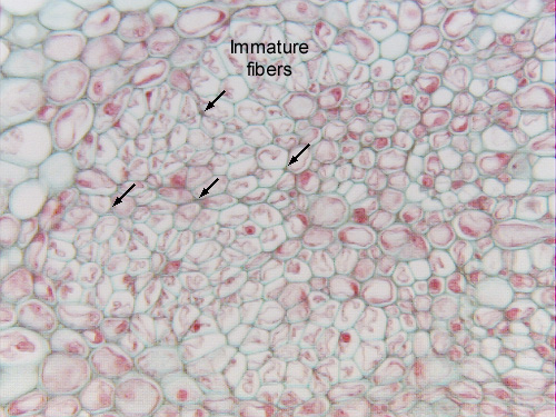
Fig.
11.5-16. Transverse section of oak stem (Quercus). This is
such a high magnification that the image is slightly pixelated. And even at this
high magnification, it is not possible to see clearly the collapsed protophloem
sieve tube members. The
arrows indicate tiny dark smudges that appear to be good candidates for
collapsed phloem.
Along the outer edges of this mass of phloem are large cells that were plasmolyzed during fixation – these are the cells that would have become the phloem fiber caps.