INFLUENZA
I. GENERAL
II. HISTORY
III. INFLUENZA VIRUSES
IV. INFLUENZA VIRUS STRUCTURE
V. ANTIGENIC DRIFT AND SHIFT
VI. CLINICAL DISEASE
VII. PATHOGENESIS
VIII. DIAGNOSIS
IX. PREVENTION
X. TREATMENT
INFLUENZA
I. General Comments
A. Respiratory Disease
Account for 75-80% of all acute morbidity in US
~80% of these illnesses are viral (average 3-4 illnesses/year/person
Frequency greatest in children
Seasonality - incidence lowest in summer and highest in winter
Major respiratory viruses involved in acute respiratory disease: influenza, parainfluenza, rhinovirus, adenoviruses, respiratory syncytial, and respiratory corona
All of these agents associated with increased risk of bacterial superinfection of the damaged tissue of the respiratory tract
All have worldwide distribution
II. History of Influenza
A. Major epidemic disease of developed countries
22 major pandemics recorded since early 18th century
Spanish influenza pandemic (1918 - 1919)
(World War I); One of most devastating plagues in history; killed 20 million and affected large part of human population
Influenza: Name from Italian form of Latin influentia: reflected widespread superstition that epidemics resulted from astrologic or occult influence such as an unhappy conjunction of the stars
Originally thought disease caused by Hemophilus influenzae (hence name)
1933- Smith, Andrews, Laidlaw found that filtered, bacteria-free nasal washings from influenza patients produced a characteristic febrile disease when inoculated intranasally into ferrets
III. Influenza Viruses (Figure 1, Figure 2)
A. Group Characteristics: Enveloped, pleomorphic, possess single stranded RNA
B. Classed into 3 major serotypes
A: Causes more severe disease and extensive epidemics
Greater tendency to undergo antigenic changes
Found in man and animals
B: More antigenically stable
Occurs in more localized outbreaks
Restricted to man
C: Rarely causes clinical disease
Not involved in epidemics
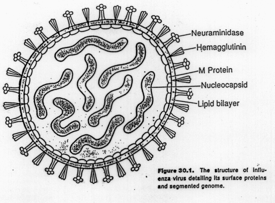
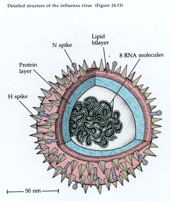
IV. Influenza A Virus Structure
Envelope of glycolipid derived from the host plasma membrane
Nucleocapsid containing segments of negative-sense, single-stranded RNA
Important surface glycoproteins:
Hemagglutinin: agglutinates certain kinds of red blood cells (e.g. chicken)
Major function - to serve in attachment to mucoprotein receptor sites on human respiratory cell surface (1st step in infection)
Antibody to HA neutralizes virus infectivity
Neuraminidase:
a. Antigenic hydrolytic enzyme
b. Acts on mucoprotein hemagglutinin receptors (splitting off terminal neuraminic acid.
c. Destruction of receptor activity (seems important for releasing virus from infected cell
Host viral receptor
Glycoproteins possessing a terminal N-acetylneuraminic acid group (NANA)
Adsorption of virus leads to release of NANA by viral neuraminidase
V. Antigenic Shift & Antigenic Drift
A. Unique aspect of Influenza A is ability to develop wide range of subtypes through mutation and recombination. These result in antigenic changes called drifts and shifts.
1. Subtypes based on H and N antigens
13 major H subtypes (H1, H2, H3 are important in human infections)
9 subtypes of neuraminidase antigens (N1, N2 are of greatest importance in human infections)
Viral subtypes designated according to the H & N antigens on their surface
DESIGNATION OF VIRAL SUBTYPES
A/Texas/1/77(H3N2)
A = virus type
Texas = city, state, or
country of first isolation
1 = Strain
77 = year of recovery (1977)
(H3N2) = Hemagglutinin &
Neuraminidase subtypes
2. Designation of influenza A subtypes: designated according to major representative virus to which they are most closely related antigenically using place of initial isolation, number of isolates, and year of detection
e.g. A/Texas/1/77(H3N2)
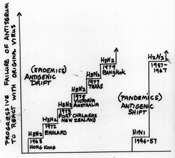
3. ANTIGENIC DRIFT: occurs as result of natural selection of mutants under selective pressure of increasing levels of immunity in population.
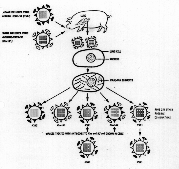
4. ANTIGENIC SHIFT: Major changes in H and N subtypes of epidemic strains can occur rapidly and unpredictably
a. May result from recombination in which different influenza subtypes simultaneously infect a cell and produce progeny that contain antigens from either of the original viruses
e.g. A(H3N2) + A (H1N1) ---> Progeny (H3N2,H1N1,H1N2,H3N1)
[Observed in lab and probably occurs in nature] (Figure 3)
b. OR certain serotypes circulate into animal or avian reservoirs only to reemerge and adapt to human hosts when a sufficient proportion of population has little immunity to “new” subtypes
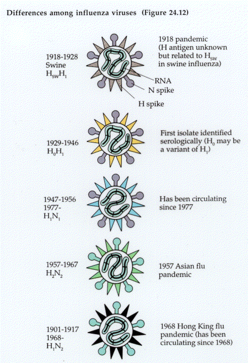
c. Major antigenic shifts (recently) have occurred approximately every 8-10 yrs. (Figure 4)
Often result in serious epidemics or pandemics
d. Eventually overall immunity of population becomes sufficient to minimize epidemic potential of major subtype and its drifting strains (Figure 5)
e. Scene set for sudden and unpredictable appearance of an entirely new subtype that may not have circulated from 20 or more years.
VI. Clinical Disease:
A. Infects upper and lower respiratory tract
B. Short incubation period: average of 2 days; onset abrupt with symptoms developing over a few hrs
Fever, Myalgia, Fatigue, Headache, Occasional shaking chills
Within 6-12 hrs patient usually at peak of severity with dry non-productive cough
Illness remains severe - sometimes with worsening cough for 2-5 days followed by gradual improvement
1 week after feel better but fatigue, nonspecific weakness and cough can linger 2-3 weeks
Complications: Bacterial superinfection-most commonly S. pneumoniae, H. influenzae, S. aureus
C. Reye's Syndrome- Infants and children 2-12 days after onset of viral infection (any of a variety of systemic viral illness). Severe hepatic dysfunction and cerebral edema
VII. Pathogenesis
A. Effects on infected cells
Virus lytic; Switch off host cell protein and nucleic synthesis
Cause release of lysosomal hydrolytic enzymes
Cell death activates complement components --> leads to localized inflammation
B. Impairment of host defenses
Functions impaired: PMN chemotaxis and phagocyte function; alveolar macrophage activity; ciliated respiratory epithelial cell function; T-lymphocyte responsiveness
C. Recovery from infection
Initially by interferon production - limits further viral replication
Generation of natural killer and cytopathic T cells
Followed by appearance of local and humoral antibody and cellular immunity --> finally tissue repair
VIII. Diagnosis
1. Viral isolation during acute phase of illness
2. Serological diagnosis (important in epidemiology) - 4-fold or greater increase in antibody titers in acute and convalescent specimens collected 10 -14 days apart
IX. Prevention
A. Killed virus vaccine prepared from those strains most closely related to antigenic subtypes causing infections.
1. May contain whole virus or “split” subunits composed of hemagglutinin antigen
2.. Vaccine efficacy variable and annual revaccination necessary for maximal protection
B. Prophylaxis
1. Amatadine hydrochloride (or Rimantadine) : acts by inhibiting viral uncoating or primary transcription of viral RNA
Recommended only for high risk cases
X. Treatment
Symptomatic care and anticipation of potential complications especially bacterial superinfection
Children should not use aspirin (Reye's)