HEPATITIS
Hepatitis is a syndrome, not a single infectious agent.
Characterized by inflammation of the liver.
At least 4 types of viral hepatitis:
Type A - infectious
Type B - serum
Type C - transfusion
Type D - delta,
Type E
NANB- nonA nonB (no viral agents have been identified)
Outcome of infection with one of these agents is extremely variable, varying from completely asymptomatic to fatal.
Can be divided into 4 outcomes:
1. Typical, acute icteric (jaundice) hepatitis
2. Subclinical, anicteric - most common
3. Fulminant hepatitis - relatively rare
4. Chronic hepatitis - hepatic inflammation and necrosis lasting for at least 6 months.
10% type B
10 - 60 of C and D.
Does not occur with type A.
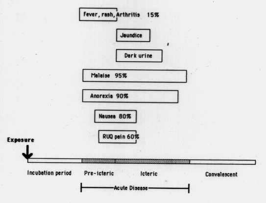
Acute viral hepatitis: course of infection separated into 4 stages (Figure 1):
1. Incubation period - average (range)
A - 25 (15 - 45)
B - 75 (40 - 1 80)
D - (30 - 50)
NANB - 50 (15 - 150)
2. Pre-icteric phase
Non - specific symptoms before the onset of topical hepatitis. Most common complaint is malaise. Usually see anorexia, often followed by nausea, vomiting.
Weight loss of 2 - 10 lbs is common. Right upper quadrant (RUQ) abdominal pain is a common complaint but is usually not severe.
About 10 - 15% develop a serum-sickness-like syndrome in the early phase, characterized by low-grade fever, rash and arthritis.
3. Icteric phase
Dark urine and/or jaundice due to increased levels of bilirubin
Disease is usually diagnosed during this period.
Biochemical tests: look for marked elevation of serum levels of the liver enzymes alanine aminotransferase (ALT) and aspartate aminotransferase (AST). In acute hepatitis, these are almost always elevated 10-fold and can be as high as 100-fold. Enzymes levels increase during late incubation period and are always elevated once symptoms appear.
Prothrombin time is usually normal or only slightly prolonged, marked prolongation indicates ervere, possibly fulminant disease. Blood counts are usually normal except in fulminant cases.
4. Convalescent phase
Begins with the disappearance of juandice and other symptoms. Enzyme levels will fall but will remain slightly elevated for some time after symptoms disappear. Serolocial tests can be used at this stage to identify the viral agent responsible. NANB usually diagnosed by exclusion.
Hepatitis Viruses
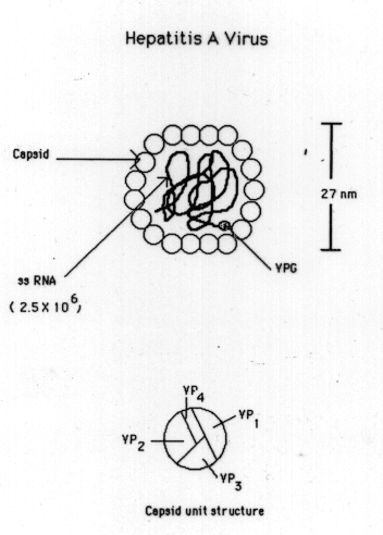
Type A (Figure 2): HAV is a small, 27-nm RNA virus that belongs to the picornavirus family. Evidence indicates that it is a simple nonenveloped virus with a nuclocapsid designated the hepatitis A antigen (HA Ag). The viral capsid consists of 32 capsomeres arranged in an icosahedral conformation. Each capsid is composed of four polypeptides (VPI - VP4). Inside the capsid is a single molecule of ssRNA approximately 8,100 nucleotides in length.
+ strand virus (positive polarity - proteins are translated directly off the RNA). The 3' end of the RNA is polyadenylated and the 5' end has a small protein, the "viral protein, genomic" (YPg) which may aid in attaching the RNA to cell ribosomes. Virus can be grown in tissue culture - replicates in the cytoplasm and is noncytopathic.
Virus is related to polio virus and has a similar epidemiology:
Spread by fecal - oral route
Spread is usually associated with overcrowding and poor hygiene
Contaminated food and water are the usual modes of transmission
Can occur as outbreaks or epidemics but also occurs sporadically in industrialized nations - accounts for ~20% of viral hepatitis in adults in the U.S.
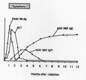
Course of hepatitis A - (Figure 3)
Incubation period is relatively short
Shedding of antigen (i.e. virus) in feces is maximal just before appearance of symptoms - very important in terms of spread
Shares some properties in common with polio:
children more often asymptomatic in areas of poor sanitation,
virus tends to be endemic,
children infected early possibly while still partially protected by maternal antibody and outcome less severe
Where children escaped early exposure - more likely to be symptomatic when infection does occur
Detection:
Hepatitis A virus is usually not detected in serum.
Antibody to HAV is detected with the onset of symptoms. IgM detected early and falls off in titer after symptoms resolve. IgG rises later and persists for life. Diagnosis of HAV usually depends on finding anti HAV IgM in the serum during the acute phase.
Disease can be severe and prolonged but only rarely fatal (0.1% of icteric cases) and does not lead to chronic carriage.
Treatment
With g globulin - antibody fraction from pooled normal human serum. significance of the protective effect: indicates large proportion of the population has had, and developed antibody against HAV.
Hepatitis B
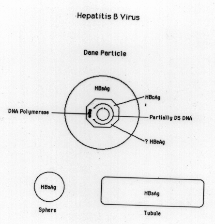
HBY is a complex, 42 nm DNA virus - unrelated to the other hepatitis viruses
Structure: (Figure 4)
Intact virus is referred to as the "Dane particle"
Composed of an outer surface component - HBsAg and an inner, core component - HBcAg inside the core is the genome - single molecule of partially double stranded DNA - single stranded region comprises 10 - 50 % of the DNA
The complete strand is ~3600 nucleotides and is nicked. This strand contains all the genetic information for producing HBsAg and HBcAg. Core contains a DNA-dependent DNApolymerase - can fill in gap. Unusual DNA structure may enhance integration of the DNA into host DNA.
(Figure 5) Serum of infected person contains HBsAg - most not Dane particles - Serum contains both 20 nm spheres and 20 nm diameter tubules (50 - 250 nm in length). These structures consists entirely of HBsAg, no HBcAg or polymerase or DNA. Concentration up to 10^13 particles/ml = 100 - 500 mg/ml average concentration. The production of large amounts of circulating antigen is a unique feature of HBV not observed with other hepatitis viruses.
HBeAg
Another Ag - HBeAg, is thought to be associated with the HBV core, circulates as a soluble serum protein, either free 19k daltons (smal e) or associated with serum immunoglobulin 300k (large e)
HBeAg is the most reliable marker for infectivity. While HBsAg positive individuals should be considered potentially infectious, correlation between HBsAg and infectivity is not uniform
HBSAg+ who are also HBeAg+ are usually highly infectious. Those without HBeAg or who have antibody to HBeAg (anti-HBe) usually have little or no circulating virus and are at less risk for spreading the disease
RIA or ELISA for HB-PAG
There are other markers for infectivity but these are more complicated assays:
1. look for Dane particles in EM
2. test for DNA polymerase by incorporation of tritiated thymidine
3. test for specific HBV DNA by molecular hybridization
Epidemiology:
Spread parenterally or by direct contact - virus is found in biological fluids, esp blood, and semen, lower in saliva. Epidemiology similar to HIV but HBV has higher infectivity.
Parenteral spread - blood products, contaminated needles (particularly among drug abusers), dialysis patients medical personnel treating high risk groups often infected by inapparent parenteral spread e.g. contamination of small breaks in the skin with blood or saliva.
Close contact - often sexually transmitted esp. among male homosexuals.
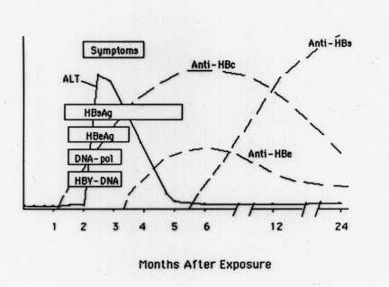
Course of disease - (Figure 6)
Incubation period: 40 - 180 days, length inversely proportional to dose.
First serologic marker to be detected is HBsAg and it persists the longest. Peaks with onset of elevated enzyme levels.
Seroconversion from HBeAg to anti-HBeAg usually associated with peak in clinical symptoms. Failure to seroconvert during acute stage is associated with progression to chronic form of the disease
Free HBcAg is not found in the serum but antibody to HBcAg appears shortly before the onset of symptoms. Ab is indicative of either ongoing or previous infection. Persistent Ab is IgG rather than IgM, which is found only during the acute phase
Ab to HBsAg differs from the other Ab's in that it appears during convalescence rather than during the acute phase. This is probably the major protective Ab - marker for recovery and immunity. HBV immune globulin (HBIG, hyperimmune globulin) is so named becuase it contains high titers of anti HBs. Much more effective in preventing hepatitis B than is gamma globulin.
Most frequent response to HBV infection is asymptomatic infection. Usually develop high titers of HBsAg antibody, sometimes in the absence of detectable HBsAg. Lasting immunity results. 1-3% of adult acute cases progress to fulminate, fatal hepatitis. Develop signs of hepatic encephalopathy: changes in behavior and sleep patterns early, Later, lethargy and finally coma ensues. Usually a decrease in hepatic size, high fever, confusion. Hepatic failure. Usually no seroconversion to anti HBeAg or HBsAg.
Vaccine
(1) HBs isolated and purified from human serum
(2) recombinant vaccine
Chronic Hepatitis:
5 - 10 of acute cases develop a chronic HBs-AG carrier state - probably persists for life.
HBe and polymerase appear in the serum at the some time as HBs. Persistence of high levels of HBeAg during the acute phase is suggestive of development of the chronic state. Highest levels of HBcAg antibody are found in the chronic carriers but little or no HBsAg antibody
As in acute hepatitis, seroconversion to anti-HBeAg is associated with a favorable prognosis.
Complications of chronic state - immune complex disease e.g. arthritis, glomerulonephritis
Also associated with primary hepatocellular carcinoma.