CHLAMYDIA
Small, obligate intracellular parasites which resemble the rickettsiae.
Two species, only share about 10% DNA homology but are thought to have arisen from a common ancestor
C. psittaci - found in a variety of vertebrates
C. trachomatis - appears to have only humans as a reservoir
Once considered to be viruses but are now known to be bacteria:
Have both DNA and RNA
Have a discrete cell wall, appear to be gram negative: peptidoglycan and an outer lipid layer
Have bacterial type ribosomes
Can synthesize proteins
Susceptible to several antibacterial antibiotics
Small genome - about 1/4 the size of the E. coli chromosome
Obligate intracellular parasitism apparently due to their inability to generate energy:
depend on host cell for ATP production and for reoxidation of reduced NADP
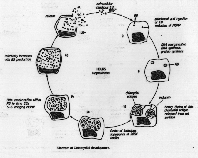
Developmental cycle:
Replication of the chlamydia involves a developmental cycle within the host cell
(Figure 1)
Elementary body - Infective particle
small, dense spherical body 0.2 - 0.4 Á in diameter
surrounded by a rigid "gram-negative like" cell wall
DNA organized into an electron dense central nucleoid
Stages in development:
1. Attachment
2. Penetration - enter the cell by a process which appears to be endocytosis, surrounded by the host cell membrane - vacuole
3. Transition from elementary body to reticulate or initial body: elementary body begins the process of reorganization within the 1st hour after infection, process requires 8 - 12 hours, nucleoid structure becomes less dense - then transcription and replication begin
Elementary body gradually enlarges ~0.7 - 1.0Á
numerous ribosomes can be seen but no discrete nucleoid
divide by binary fission
Reticulate bodies use host ATP - require functioning mitochondria in host cell, also use host purine and pyrimidine pools but biosynthesis of the reticulate body, membrane is encoded by the chlamydia
By 24 hours, the host cell nucleus has been displaced by reticulate bodies
C. trachomatis - synthesizes large amounts of glycogen which surrounds the chlamydia within the vacuole = inclusion body visible on histologic staining
C. psittici - more diffuse, no glycogen, membrane ruptures leaving the chlamydia in the cytoplasm
4. Maturation:
By ~30h post infection, the reticulate bodies reorganize into elementary bodies and synthesize the elementary body cell wall, increase in density and decrease in size
Various stages of development can be seen within the same cell at this stage
5. Cell ruptures and releases infective elementary bodies.
Cultivation:
Cannot be grown on artificial media, can grow in
1. chick embryo yolk sac
2. Tissue culture cells - HeLa cells or McCoy mouse cells, usually necessary to irradiate or treat the tissue culture cells with cyclohexamide to inhibit host replication and enhance chlamydial growth
3. Some strains will infect mice
Antigenic structure
C. trachomatis and C. psittaci share a common group antigen - LPS - not protective
Protective antigens are type specific cell wall antigens
DISEASE
| Species | Serotype | Disease | 1║ Reservoir | Distribution |
| C. psittaci
|
many
|
Psittacosis-febrile disease,influenza like symptoms +/- pneumonia |
Birds
|
Worldwide
|
| C. trachomatis
|
A, B, C
|
Trachoma - eye disease
|
man
|
1║ endemic in Asia & Africa |
| D, E, F, G, H, I, J, K, L, M
|
Genital trachoma NGU, cervicitis, endometritis, salpingitis Inclusion conjunctivitis Infant pneumonia
|
man
|
Worldwide
|
|
| LGV L1, L2, L3 | Lymphogranuloma venereum | man | Worldwide | |
| C. pneumniae | Respiratory syndromes | man | Worldwide |
Transmission -
Organisms are not hardy, do not survive long outside the body
Transmission requires close contact, or in the case of C. psittici, rapid spread by dust or aerosol
Pathogenic mechanisms -
1. Compete with host cell for nutrients - cell death and tissue damage
2. When ingested by phagocytic cell, non-opsonized chlamydia prevent lysosomal fusion with the phagacytic vacuole (opsonized cells are more easily killed by macrophages)
3. All sppecies produce serotype specific heat labile protein toxins that are lethal when injected IV into mice
Immunity -
Protection requires both Ab and CMI - nude mice are more susceptible and Ab give only partial protection
Psittocosis
Zoonosis - contracted by inhalation of respiratory secretions or dust from dropping of infected birds. Disease is usually latent in the natural host (bird) - activated under stress, e.g. recent captivity or transport
Wide variety of avian species can be infected:
Occupational hazard of poultry workers, esp. turkeys, incidence reduced by inclusion of drugs in feed
Pet psittacine birds ( parrots and parakeets, ets) - reduced by quarentine regulations for imported birds
Develops as an acute infection of the lower respiratory tract -
fever, headache, malaise, muscle aches, cough, X-ray reveals a bilateral interstitial pneumonia
may lead to myocarditis, encephalitis, and/or hepatitis
patient usually gives a history of bird exposure - can be acquired from symptomatic or asymptomatic birds
Diagnosis - 4X rise in titer against the group Ag, difficult to isolate the organism and hazardous to lab personnel
probably a large number of asymptomatic infection in humans since antibodies are found in people with no known history of the disease
Pathology -
affected tissues - liver, spleen - focal necrosis, predominance of mononuclear cells - implies CMI
lung - monnuclear cells, exudate
C. trachomatis
Trachoma - restricted to human - leading cause of blindness worldwide. American Indians in U. S. -
chronic infection often leading to blindness
(Inclusion conjuctivitis is a similar disease of adults and newborns but is not associated with chronicity or permanent eye damage)
1║ exposure usually acquired in infancy or early childhood from mother or other close contact- acute conjuctivitis - usually resolves
Persistence of the original infection or reinfection leadsd to inflammatory response - chronic inflammation with increased vascularization of corneal conjuctiva - scarring of cornea and abmormal conjunctiva - loss of vision occurs 15 - 20 years after the initial infection
No apparent immunity develops
Disease associated with poor hygiene
Genital tract infections
Similar spectrum of diseases as N. gonorrhoeae - epidemiology very similar
About 40% of case of NGU - total number of cases may exceed those due to N. gonorrhoeae
Highly prevalent: 5-10% of private patients in for prenatal exams are infected
About 50% of infants born to mothers excreting C. trachomatis during labor will develop disease -
usually inclusion conjuctivitis but 5 - 10% develop pneumonia, rarely fatal but usually a prolonged illness
Lymphogranuloma venerum is a distinct sexually transmitted disease associated with specific serotypes - relatively rare in North America
Begins as a small, painless, genital ulcer -2 to 6 weeks - swelling of regional lymph nodes - suppurative, often with a draining sinus
May cause ulcerative colitis in homosexual males
Chronic, may remain latent in some cases.